Scoliosis
Spinal column disorders can occur on two planes: the medial, where lordosis and kyphosis are observed, and the frontal, where scoliosis is evident. Scoliosis is a highly-complex spinal deformity characterised by two clear aspects: lateral curvature and vertebral rotation.
Causes of Scoliosis
The onset and progression of the deformity usually goes unnoticed, starting in puberty and affecting mainly female patients, and it's origin, in most cases, is unknown, for which it is referred to as idiopathic scoliosis. The direction of the scoliosis, either right or left, is defined by the convexity of the curvature, and its location is determined by the vertebra that is most deviated laterally from the midline, known as the apical or apex vertebra. This vertebral rotation produces a thoracic deformity because, as the vertebra rotates, the ribs on the side of the convexity are pushed back, resulting in greater prominence in the back, known as gibbosity. Other disorders such as vertebral wedging - the narrowing of the vertebral body on its concave side or widening on its convex side - are visible along with variations in the thickness and elongation of the plates. Depending on their degree of flexibility, two types of scoliosis can be discerned: structural and nonstructural, the latter of which has a higher degree of flexibility. Idiopathic scoliosis, which is of unknown origin, is the most common.
Symptoms of Scoliosis
In the case of infantile, juvenile and adolescent idiopathic scoliosis, onset and progression usually go unnoticed. They can be distinguished in lumbar scoliosis, which is common in girls, whose apex is found from T11 to L5, and are curves of a few degrees and accompanied by compensation curves. Thoracic curves are mostly right convexity, with no signs of discomfort, most commonly located between T5 and T12 and accompanied by compensation curves. Radiological examination, among others, provides information to determine the severity of the curve, the Risser sign, vertebral rotation and Cobb angle, along with other factors such as age or origin (neurofibromatosis, idiopathic, etc.) that enable full assessment of the scoliotic curve. This enables classification according to degree of deviation: 1. 0° - 20° COBB • 2. 21° - 30° COBB • 3. 31° - 50° COBB • 4. 51° - 75° COBB • 5. 76° - 100° COBB • 6. 101° - 125° COBB • 7. 126° - > COBB. Vertebral rotation, depending on the position of the pedicles, shows if there is 0° rotation or, conversely, 1°, 2°, 3° or 4° rotations, taking as a reference the pedicle of the convexity with respect to the vertebral edge. Other factors, such as bone maturity, determine the type of treatment required.
Scoliosis Braces ~ Orthotic Treatment
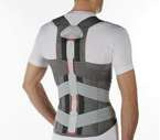
Depending on factors such as pain, origin and etiology, degree of deviation, patient age, etc., treatment can be conservative, rehabilitational or surgical. Braces for the treatment of scoliosis can be active (corrective action) or passive (stabilisers and/or immobilisers). Cases of juvenile and adolescent idiopathic scoliosis, whose curves are lumbar, dorsal or dorsolumbar, and in a Cobb angle range of between 20° and 50° with varying degrees of vertebral rotation, require orthotic treatment with active braces. Throughout the history of scoliosis treatment, different types of brace have been designed, with the Boston brace being the most used today. Developed by John Hall and Billy Millar, its current design is still based on the original Boston brace for the quality of its properties, such as the materials used, components and the morphology of the module. In the case of high dorsal or dorsolumbar curves, treatment can be carried out with the tlso braces. Cotrel night and/or daytime auto-traction systems are used by some interdisciplinary teams as a way to provide more pre-intervention elasticity by reducing the Cobb angle to a certain extent and observing the curve’s degree of elasticity. This orthopedic support is created from a prefabricated polypropylene module with an interior Naturplast lining and has a wide range of sizes and lumbar lordosis correction options to enable the patient to select the right size according to a table of specific measurements for each patient. Some patients with peculiar morphologies may require the manufacture of a specific made-to-measure module. The design, trim, appearance of the decompression windows and location of the corrective wedges for a custom design are determined by the curve to be treated and the relevant vertebral segment. Its correction principles are based on a set of forces applied to the convexity of the curve, seeking compression and derotation of the vertebrae, which involves correct and close positioning of the corrective wedges and proper location of the expansion windows.


